The ELISA test: Enzyme Linked Immnoasorbent Assay
For this test we tested the presence of antibodies. In this process we add 50 micro liters of purified antigen marked AG into each of the 6 wells marked positive, negative, 8 and 47. After letting it sit for 5 minutes to allow the proteins to bind to the plastic wells via hydrophobic interaction. Then we washed it out using a wash buffer and a plastic pipet.


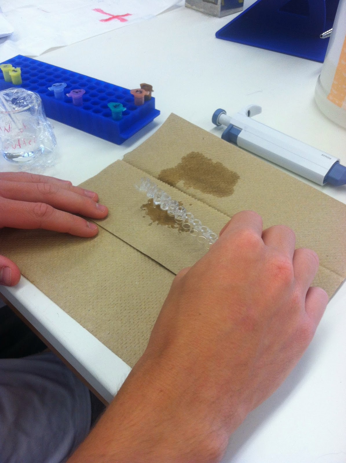
We added the serum samples and control samples. The positive control in the violet tube was added to the two positive wells, the negative control in the blue tube was added to the two negative wells and the each serum sample was added to the marked wells (8 and 47). Then we let it sit for 5 minutes. This serum that we added contains millions of different type of antibodies, but only if the serum contains the right antibodies for the virus antigen will the antibodies bind to the antigen in the wells


After dumping out the serum and control sample we washed out the wells using the wash buffer. Then we added the 50 microliters of the secondary serum marked SA in the orange tube. This will detect the serum or primary antibodies. For if the serum antibodies have bound to the antigen, the secondary antibodies will bind tightly to the serum antibodies. We waited 5 minutes and then dumped out the secondary serum and the washed it our using the wash buffer.


Now the secondary serum is bound to the enzyme (HRP) that chemically changes the enzyme substrate which will turn it blue or remain colorless depending on the presence of the antibody. To see these results we added 50 microliters of the enzyme substrate marked SUB in the brown tube. If the primary antibody is present in our serum the wells will turn blue and signify a positive test. If it remains colorless the primary antibody was not present in the serum, a negative test.
Our results were positive for the first two wells of positive control. The negative control wells were negative. The well marked 47 was also negative, however, due to slight contamination, it turned blueish later on. The well marked 8 was positive as it turned blue initially.


Antibody-Antigen Reaction in Agar and Food Purity Follow Up::
After allowing the plate to sit at room temperature for a day, we observed the results of the antibody-antigen reaction and food purity tests. A cloudy white strip can be seen between the bovine albumin and the goat anti-bovine albumin. Naturally, this would not occur with the goat anti-swine or goat anti-horse albumin because the bovine antigen would not react with a horse or swine antibody.

The same result happened in the food purity test where a white strip is shown between the well with hamburger extract and the well containing the goat anti-bovine albumin. This means that the hamburger extract only contains bovine albumin. If a white strip were to appear between the hamburger extract and either the anti-horse or anti-swine albumin, then that would signify a contamination that may have occurred during meat processing.

Yogurt:
We also made yogurt, and tested the effects of using no heat and heated milk. After heating the milk to a boil and letting it cool down to 37 degrees. Placing it in Styrofoam cups Dr. P inoculated the milk by adding small spoonful of Yoplait yogurt into each cup. (Unfortunately this yogurt did not have as many active strains so we don’t expect optimal results) Then we placed them in the incubators and lety the yogurt proteins work on the unheated and heated milk.


CAMP test:
Because we were unsure about Elizabeth’s result of strephtolococcus aureas from her throat swab test on Blood agar, Dr. P did an addition test called the CAMP test. This test is to test beta-hemolysis caused by strephtolococcus aureus. Three inoculations are on the test and if there is an arrow present this indicated a positive test.
No comments:
Post a Comment