Friday, May 30, 2014

Day 12: 5/30/14

Discovering our Unknown Bacteria “N”

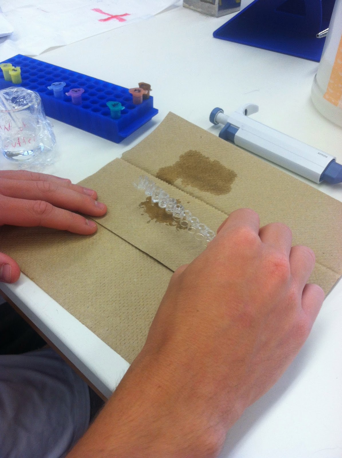
Today we discovered what our bacteria is. So by using a flow chart we deduced this:

Our bacteria consists of Gram negative facultative anaerobic rods.


The oxidase test turned out negative- it does not have cytocrome oxidase because no electrons were added to the oxidase reagent. Therefore it is an Enterobacteriaceace.


Our bacteria was a strong lactose fermentor- the test proved gas production during lactose fermentation.  It also had a negative test for Indole or tryptophan- our bacteria cannot split tryptophan into indole and pyruvic acid. Because it cannot break down tryotophan it does not use it as a source of energy.


The MRVP test was negative for methyl red and positive for Voges-Proskeur was positive. Methyl red test was negative meaning our bacteria cannot ferment glucose via mixed acid fermentation. The Vogues test showed our bacteria however can ferment glucose.


Now the choices of our bacteria were narrowed down to two ones E. aerogenes and E. cloacae. After battling it out against Peter and Ben we finally claimed E. cloacae as our bacteria and here’s how:
-Our rods are very small- 0.5 to 1.0 to 1.0 to 2.0 microns (as opposed to just small rods for E. aerogenes)
-During fermentation of sugars produced acid and gas actively from glucose and sucrose and less actively from lactose


-Our test was positive for nitrate (meaning our bacteria has a nitrate reductase enzyme)
-Although both of the bacterias were motile, our proved to be more motile than the other bacteria.


-and finally the temperature growth. Our bacteria grew optimally at 30 degrees Celsius.  During the course of the lab it also grew at 37 degrees Celsius when we shared plates with another group. According to the book the temperature for E. cloacae is between 30-37 degrees Celsius.
-the other bacteria E. aerogenes also grows at 30 degrees Celsius, but can also grow at lower temperatures. Peter and Ben’s bacteria grew at 25 degrees Celsius. Therefore after much research and debate, there’s turned out to be E. aerogenes.
FINAL RESULT: Enterobacter clocae
Habitat: in human and other feces and in sewage, soil and water.

Yogurt: 
After letting our yogurt sit for a day we made a fresh yummy yogurt! Because of the active cultures (although there was only one strand) added from the Yoplait yogurt the bacteria grew and multiplied in the milk to form yogurt. Heating the milk beforehand had killed any competing bacteria in the milk and therefore the heated milk had a better result. Delicious!


CAMP: Unfortunately the CAMP test was melted underneath the light, but there was an arrow indicating a reaction and testing positive for S. aureas in Elizabeth.

Well it’s been real, Hope you have enjoyed our blog! Thanks for reading, we had a blast during lab and now our lonely bacteria is no longer unknown! Mission accomplished.

Day 11: 5/29

The ELISA test: Enzyme Linked Immnoasorbent Assay
For this test we tested the presence of antibodies. In this process we add 50 micro liters of purified antigen marked AG into each of the 6 wells marked positive, negative, 8 and 47. After letting it sit for 5 minutes to allow the proteins to bind to the plastic wells via hydrophobic interaction. Then we washed it out using a wash buffer and a plastic pipet.  


We added the serum samples and control samples.  The positive control in the violet tube was added to the two positive wells, the negative control in the blue tube was added to the two negative wells and the each serum sample was added to the marked wells (8 and 47). Then we let it sit for 5 minutes. This serum that we added contains millions of different type of antibodies, but only if the serum contains the right antibodies for the virus antigen will the antibodies bind to the antigen in the wells


After dumping out the serum and control sample we washed out the wells using the wash buffer.  Then we added the 50 microliters of the secondary serum marked SA in the orange tube. This will detect the serum or primary antibodies. For if the serum antibodies have bound to the antigen, the secondary antibodies will bind tightly to the serum antibodies. We waited 5 minutes and then dumped out the secondary serum and the washed it our using the wash buffer.  


Now the secondary serum is bound to the enzyme (HRP) that chemically changes the enzyme substrate which will turn it blue or remain colorless depending on the presence of the antibody.  To see these results we added 50 microliters of the enzyme substrate marked SUB in the brown tube. If the primary antibody is present in our serum the wells will turn blue and signify a positive test. If it remains colorless the primary antibody was not present in the serum, a negative test.
Our results were positive for the first two wells of positive control.  The negative control wells were negative. The well marked 47 was also negative, however, due to slight contamination, it turned blueish later on. The well marked 8 was positive as it turned blue initially.


Antibody-Antigen Reaction in Agar and Food Purity Follow Up::

After allowing the plate to sit at room temperature for a day, we observed the results of the antibody-antigen reaction and food purity tests. A cloudy white strip can be seen between the bovine albumin and the goat anti-bovine albumin. Naturally, this would not occur with the goat anti-swine or goat anti-horse albumin because the bovine antigen would not react with a horse or swine antibody.


The same result happened in the food purity test where a white strip is shown between the well with hamburger extract and the well containing the goat anti-bovine albumin. This means that the hamburger extract only contains bovine albumin. If a white strip were to appear between the hamburger extract and either the anti-horse or anti-swine albumin, then that would signify a contamination that may have occurred during meat processing.


Yogurt: 
We also made yogurt, and tested the effects of using no heat and heated milk.  After heating the milk to a boil and letting it cool down to 37 degrees.  Placing it in Styrofoam cups Dr. P inoculated the milk by adding small spoonful of Yoplait yogurt into each cup. (Unfortunately this yogurt did not have as many active strains so we don’t expect optimal results) Then we placed them in the incubators and lety the yogurt proteins work on the unheated and heated milk.


CAMP test:

Because we were unsure about Elizabeth’s result of strephtolococcus aureas from her throat swab test on Blood agar, Dr. P did an addition test called the CAMP test. This test is to test beta-hemolysis caused by strephtolococcus aureus. Three inoculations are on the test and if there is an arrow present this indicated a positive test.


Thursday, May 22, 2014

Day 5: 5/20

Follow up On Anaerobic Bacteria and Motility Test and Start of Food Test:

After a lovely game of frisbee we returned to the lab today to follow up on the experiments we started yesterday.  The first was the Thiglutinate test.  Our results were a bacteria that grew all the way through both the oxygenated and deoxygenated substance. This meant that our bacteria is facilitated anaerobic, meaning it can grow in either environments.


To follow up the the aerobic test we checked on the airtight container of or agar plates.  Unfortunately some oxygen was let into the container, and alas strip did not turn white and remained blue.  But still we could see how the bacteria grew with minimal oxygen overnight.  Our results also showed that our bacteria is anaerobic facilitated, as you can see by the growth where the inoculating tube was traced over the surface.


Today we did another trial run of the air tight container with the GasPak.


Also we checked our motility test tube.  Our bacteria was motile and the cloudiness around the inoculation lined showed the mobility of the bacteria.


Then we did a Catalase test by pouring hydrogen peroxide onto our very own plate (yay for being the odd ones out!) Because our bacteria is facilitated anaerobic there was a slight formation of bubbles meaning there was little catalase in our bacteria. This is the case because only aerobic organisms produce catalase to break down hydrogen peroxide.  Other group had more bubbles because their bacteria were anaerobe, ours had bubbles because it is facilitated anaerobic, meaning it can also grow in oxygen and does contain small amounts of catalase.


Finally we tested 10 tubes and plates for the food our bacteria eats to narrow down the type of unknown bacteria we have. Using aseptic technique we inoculated all of the tubes and plate with the following materials:
Bunsen burner
inoculating loop and needle
plate of unknown "N" bacteria


First we inoculated the starch plate, the yellow skim milk plate, DNA plate and the blue lipid plate using aseptic technique. We traced a squiggly line along on the plates.


Next we inoculated the the TSI slant tube. Sticking the inoculating needle three fourths of the way into the agar then pulled the needle out and then traced the tip along the top of the agar while pulling the needle out. We also inoculated the nutrient gelatin deep tube with three stabs with the inoculating needle.


Finally using the inoculating loop we inoculated the broth tubes of litmus milk, sucrose, lactose, maltose and glucose. Then we labeled all the tubes and plates (some many tubes and plates) and placed then on 30 degrees Celsius to grow.  This will test which nutrient the bacteria uses for growth.


Results of the virus with the Unknown Bacteria:
We got out our agar plate with the virus drawn over the inoculated tube.  The bacteria had grown everywhere and the virus had killed the areas where it was traced over the surface. We put a star and our initials E.T (I mean, we are the odd ones out, right?)


Strep Test:

And last but not least we did a strep test on Theresa (gross, gag reflex :/) After taking a swab of the back of her tongue Catherine inoculated an agar plate and then put and antibacterial agent on the plate to test for streptococcus bacteria. And tomorrow we shall see if Theresa is truly and invalid!
                 

Monday, May 19, 2014

Day 4: 5/19


Slants and Plates: The Start of Many Experiments

Mary Rose is sick:

To start off this Monday afternoon in the lab we examined the petri dish of Mary Rose's streptococcus that had grown over the weekend. We examined the dish that had bacitracin and concluded that it was in fact streptococcus (Congratulations to Mary Rose for saving 80 bucks!)


The second plate contained spots of Penicillin, cloves and cinnamon of Pete's doing.  Apparently cinnamon has been a wonder drug all along and was most effective in killing the bacteria and here we thought it was just a nice spice. Cloves were not as effective. but that is because they are used more as a topical application to the throat.


We inoculated several tubes and plates:
The materials we used were:
Bunsen burner
loop and stab inoculators
striker
China marker
2 slant tubes
1 agar deep tube of motility medium
2 petri dishes
perti dish with grown "N" bacteria
thioglycollate broth tube


Next using Aseptic technique, we inoculated two slant tubes with the unknown bacteria "N" using a stab inoculator, made three deep stabs into the agar and after marking the tubes "N" we placed them in the incubator of 30 degrees Celsius. (our other slant tubes were just a liquid disaster)


We also inoculated an agar deep tube of motility test medium for a motility test , using the Aseptic technique. Again we used the stab innoculator and after inoculating it with three deep stabs we marked the tube "N" and placed it in 30 degrees Celsius incubator.


Culturing Anaerobic Bacteria:

For this experiment our purpose was to cultivate bacteria that cannot survive in the presence of oxygen. There were two parts to this experiment, the first was to inoculate a thioglycollate broth tube with the unknown bacteria and the inoculating loop.  We made a dip with the loop with the bacteria deep into the broth tube which contained an oxygen part and a part with no oxygen. After marking it we placed it in the 30 degree Celsius incubator to grow. After 48 hours we will examine the tube for growth


The second part of this test involved a perti dish with inoculated unknown bacteria and a GasPak Anaerobic system.  After a lovely presentation by Dr. P we learned about the GasPak and how it works. This system uses a chemical reaction to consume free oxygen gas in a sealed jar.  The chemicals in the "scalding hot" envelope activates with water and eventually produces carbon dioxide which stimulates the growth of some anaerobes.  We placed a indicator strip that had turned white to blue after we opened it (yay real oxygen is in the Steubenville air!) and we placed in in the jar.  When the indicator strip turns white again we will know that there is no more oxygen in the sealed jar.  We placed our whole inoculated agar plate with the unknown bacteria into the jar and the mighty strong Pete sealed it for us.  Now, we wait for anaerobic growth.


Finally we inoculated a perti dish of agar with the streptococcus from Mary Rose and made a spread plate. We used a 50 micro liter dispenser to place the streptococcus onto the agar and then spread it out on the loop.  Then we placed a bacteriophage onto the petri dish and traced our initials and drew a star :) Next lab we should see the bacteria growing but not where the virus has been placed. Then we placed it in the 37 degree Celsius incubator because the bacteria grows in humans.