Friday, May 30, 2014

Day 12: 5/30/14

Discovering our Unknown Bacteria “N”

Today we discovered what our bacteria is. So by using a flow chart we deduced this:

Our bacteria consists of Gram negative facultative anaerobic rods.


The oxidase test turned out negative- it does not have cytocrome oxidase because no electrons were added to the oxidase reagent. Therefore it is an Enterobacteriaceace.


Our bacteria was a strong lactose fermentor- the test proved gas production during lactose fermentation.  It also had a negative test for Indole or tryptophan- our bacteria cannot split tryptophan into indole and pyruvic acid. Because it cannot break down tryotophan it does not use it as a source of energy.


The MRVP test was negative for methyl red and positive for Voges-Proskeur was positive. Methyl red test was negative meaning our bacteria cannot ferment glucose via mixed acid fermentation. The Vogues test showed our bacteria however can ferment glucose.


Now the choices of our bacteria were narrowed down to two ones E. aerogenes and E. cloacae. After battling it out against Peter and Ben we finally claimed E. cloacae as our bacteria and here’s how:
-Our rods are very small- 0.5 to 1.0 to 1.0 to 2.0 microns (as opposed to just small rods for E. aerogenes)
-During fermentation of sugars produced acid and gas actively from glucose and sucrose and less actively from lactose


-Our test was positive for nitrate (meaning our bacteria has a nitrate reductase enzyme)
-Although both of the bacterias were motile, our proved to be more motile than the other bacteria.


-and finally the temperature growth. Our bacteria grew optimally at 30 degrees Celsius.  During the course of the lab it also grew at 37 degrees Celsius when we shared plates with another group. According to the book the temperature for E. cloacae is between 30-37 degrees Celsius.
-the other bacteria E. aerogenes also grows at 30 degrees Celsius, but can also grow at lower temperatures. Peter and Ben’s bacteria grew at 25 degrees Celsius. Therefore after much research and debate, there’s turned out to be E. aerogenes.
FINAL RESULT: Enterobacter clocae
Habitat: in human and other feces and in sewage, soil and water.

Yogurt: 
After letting our yogurt sit for a day we made a fresh yummy yogurt! Because of the active cultures (although there was only one strand) added from the Yoplait yogurt the bacteria grew and multiplied in the milk to form yogurt. Heating the milk beforehand had killed any competing bacteria in the milk and therefore the heated milk had a better result. Delicious!


CAMP: Unfortunately the CAMP test was melted underneath the light, but there was an arrow indicating a reaction and testing positive for S. aureas in Elizabeth.

Well it’s been real, Hope you have enjoyed our blog! Thanks for reading, we had a blast during lab and now our lonely bacteria is no longer unknown! Mission accomplished.

Day 11: 5/29

The ELISA test: Enzyme Linked Immnoasorbent Assay
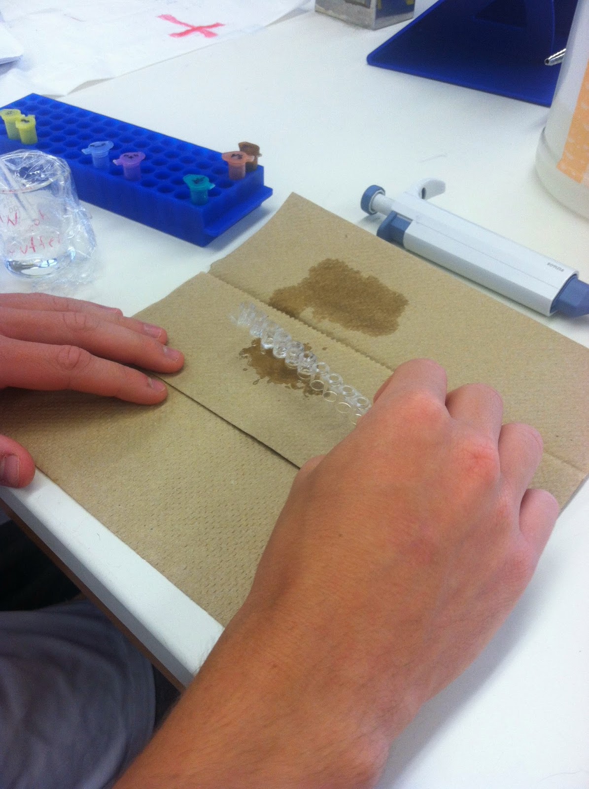
For this test we tested the presence of antibodies. In this process we add 50 micro liters of purified antigen marked AG into each of the 6 wells marked positive, negative, 8 and 47. After letting it sit for 5 minutes to allow the proteins to bind to the plastic wells via hydrophobic interaction. Then we washed it out using a wash buffer and a plastic pipet.  


We added the serum samples and control samples.  The positive control in the violet tube was added to the two positive wells, the negative control in the blue tube was added to the two negative wells and the each serum sample was added to the marked wells (8 and 47). Then we let it sit for 5 minutes. This serum that we added contains millions of different type of antibodies, but only if the serum contains the right antibodies for the virus antigen will the antibodies bind to the antigen in the wells


After dumping out the serum and control sample we washed out the wells using the wash buffer.  Then we added the 50 microliters of the secondary serum marked SA in the orange tube. This will detect the serum or primary antibodies. For if the serum antibodies have bound to the antigen, the secondary antibodies will bind tightly to the serum antibodies. We waited 5 minutes and then dumped out the secondary serum and the washed it our using the wash buffer.  


Now the secondary serum is bound to the enzyme (HRP) that chemically changes the enzyme substrate which will turn it blue or remain colorless depending on the presence of the antibody.  To see these results we added 50 microliters of the enzyme substrate marked SUB in the brown tube. If the primary antibody is present in our serum the wells will turn blue and signify a positive test. If it remains colorless the primary antibody was not present in the serum, a negative test.
Our results were positive for the first two wells of positive control.  The negative control wells were negative. The well marked 47 was also negative, however, due to slight contamination, it turned blueish later on. The well marked 8 was positive as it turned blue initially.


Antibody-Antigen Reaction in Agar and Food Purity Follow Up::

After allowing the plate to sit at room temperature for a day, we observed the results of the antibody-antigen reaction and food purity tests. A cloudy white strip can be seen between the bovine albumin and the goat anti-bovine albumin. Naturally, this would not occur with the goat anti-swine or goat anti-horse albumin because the bovine antigen would not react with a horse or swine antibody.


The same result happened in the food purity test where a white strip is shown between the well with hamburger extract and the well containing the goat anti-bovine albumin. This means that the hamburger extract only contains bovine albumin. If a white strip were to appear between the hamburger extract and either the anti-horse or anti-swine albumin, then that would signify a contamination that may have occurred during meat processing.


Yogurt: 
We also made yogurt, and tested the effects of using no heat and heated milk.  After heating the milk to a boil and letting it cool down to 37 degrees.  Placing it in Styrofoam cups Dr. P inoculated the milk by adding small spoonful of Yoplait yogurt into each cup. (Unfortunately this yogurt did not have as many active strains so we don’t expect optimal results) Then we placed them in the incubators and lety the yogurt proteins work on the unheated and heated milk.


CAMP test:

Because we were unsure about Elizabeth’s result of strephtolococcus aureas from her throat swab test on Blood agar, Dr. P did an addition test called the CAMP test. This test is to test beta-hemolysis caused by strephtolococcus aureus. Three inoculations are on the test and if there is an arrow present this indicated a positive test.


Wednesday, May 28, 2014

Day 10: 5/28/14

Follow up on the Kirby-Bauer Test:
Today we checked the results of the antibiotic test by measuring the effects of the antibodies on our unknown bacteria. Using a ruler with millimeters we measured each disc of the antibiotic. 

1. The penicillin had no effect on our bacteria and therefore is resistant (this is because our bacteria is a gram negative and has a small cell wall, so the penicillin that acts on the cell wall is not effective in killing our bacteria
2. Tetracyclin did work and measured 18mm, meaning it was in the intermediate range, our bacteria was not completely resistant but it was definitely sensitive. 
3. Neomysin was also effective and measured 17mm and was also intermediate.
*Both Neomysin and Tetracyclin work against the protein synthesis of our bacteria
4. Cinnamon was effective against the bacteria and measured 19mm
5. Lemon had no effect and the bacteria is resistant
6. The Augmentin was resistant and only measures 10mm, it did not effectively work against our bacteria


The Differential plates:
Urine Sample-
EMB plate was used to detect a strong, weak or nonlactose fermenter in coliforms. The small colony growth and pinkish tinge indicated that our sample was a weak lactose fermenter and a positive test.  Our test actually was a positive due to contamination from skin bacteria, we actually learned about in class today.  Although the sample was contaminated we learned the direct effects that everyday microflora living on our skin can have on samples.  The colony count was 4 per the 50 microliters so for every liter there would be 800 colonies.  A real positive test of a weak fermenter would have determined it was a staphylococcus. ( a strong fermenter would have meant an E. coli)  Lesson of the day: Aseptic technique is essential to microbial testing!!

Nose Sample:
For this we used a Mannitol plate to detect pH and halophiles in the nose sample. This plate also differentiates between the halophile staphylococcus aurelis (fermenter) and epididimis (nonfermentor). If MRSA was present the plate would have turned a yellow due to the pH change.  Our test results were negative. 


Throat Swab:
For the throat swab we used a Blood agar plate to look for Strephtolococcus pyrogenes or Strephtolococcus alactica.  The results of the trhaot sample were gamma hemolysis, meaning there was no lyses of the red blood cells and our test is negative.  If we had a positive result of beta-hemolysis we could have further tested for Strephylococcus pyrogenes with Bacitracin and the other groups went onto test their beta hemolysis results.  If their bacteria is sensitive to Bacitracin it will signify a positive test for S. pyrogenes


PCR – Polymerase Chain Reaction
We learned about the polymerase chain reaction test used to amplify a specific region of a DNA strand (the DNA target) for detecting antibodies and antigens and therefore a bacteria or virus. The PCR carries out this reaction using several small tubes and a thermal cycler to heat and cool the liquid during the different stages of the test.  The heating breaks apart the DNA of the bacteria or virus and adds a new complimentary substrate or strand.  Then after another series of cooling and heating and adding an enzyme, the DNA begins to replicate the new strand and the virus or bacteria can now be detected using software and graphs showing the antigens and antibodies of the DNA.


Antibody-Antigen Reaction in Agar and Food Purity
This process was to study the technique of immunology and apply it in a test for food purity.  First we made four wells in the agar after placing it over the diagram II of our handout.  We made the hole using a droping pipet. Then in each hole we placed a drop of diferent solution in each hole- Bovine Albumin, Goat Anti-horse, Anti-bovine and Anti-swine Albumin.


Then we made four more holes using template III and this time in each hole we placed a solution of either Hamburger extract, Goat Anti-horse, Anti-bovine and Anti-swine Albumin. This is to test the purity of the hamburger extract.


Finally we placed the agar plate in our very own draw and will see the results tomorrow.


Tuesday, May 27, 2014

Day 9: 5/27

MR-VP Test:

Today we kicked off Lab by redoing the MR-VP test.  Due to our blunder of the following lab we had to reinnoculate a tube of broth for these two tests. We separated the broth into two tubes using a pipet for the two tests.


For the Methyl Red test (MR) we added 5 drops of methyl red into one of the tubes.  Immediately we observed and the results were negative, signaled by a yellow-orangish color of the broth.  This test was to determine the ability of our bacteria to ferment glucose via mixed acid fermentation. These acids lower the pH of the medium to below 5 and when the methyl red was added it was changed in our broth because the pH was higher due to lack of acid fermentation and less acid is present, therefore our broth turned yellow.  Our test was negative.


For Part two of this test, the Vogues-Prokauer test,  we tested to see how our bacteria ferments glucose via buntanediol fermentation. To the second tube we added 15 drops of Barrit's reagent A ( 1% alpha-naphtol in ethanol) and 5 drops of Baritt's reagent B (40% KOH).


These two reagents will react with acetone produced during glucose fermentation in the presence of oxygen and form a red precipitate. A red color means a positive test.  After waiting thirty minutes our test was positive, meaning our bacteria uses butanediol during glucose fermentation.


Swabs and Urine:

Next we did a Throat Swab, Nasal nares swab, and  Urine sample
For the Throat swab Eleni used a tongue depressor and a sterol cotton swab and touch the back of Theresa's throat (touching the uvula and avoiding contact with the tongue) and then she inoculated a Blood agar plate to test for Streptococcus aurelis (maybe Theresa still has it? we'll find out)


Theresa's Previous Throat Culture Sample
Then for the Nasal nares swab Theres took a cotton swab and after dipping it in saline solution because a dry swab would not be effective.  Then Theresa inoculated a Mannitol salt plate to test for MRSA.


Finally Eleni graciously supplied us with a urine sample and using a 50 microliter pipet placed the urine onto a EMB sample.  Using a sterilized loop (thanks to the trusty Bunsen Burner) spread the urine over the plate.


We placed all three of these plates into the 37 Celsius incubator to see what unfriendly microorganisms are growing in our friends.

Kirby-Bauer Test:

This test was to see the results of antibiotics and home remedies on our bacteria
After inoculating a nutrient agar plate with the still unknown bacteria "N"


We added the following antibiotics to the nutrient agar plate:
1. Penicillin (which effects the cell wall)
2. Tetracycline (protein synthesis)
3. Neomycin (protein synthesis)
4. Cinnamon oil (yum)
5. Lemon oil (also yum)
6. Augmentin

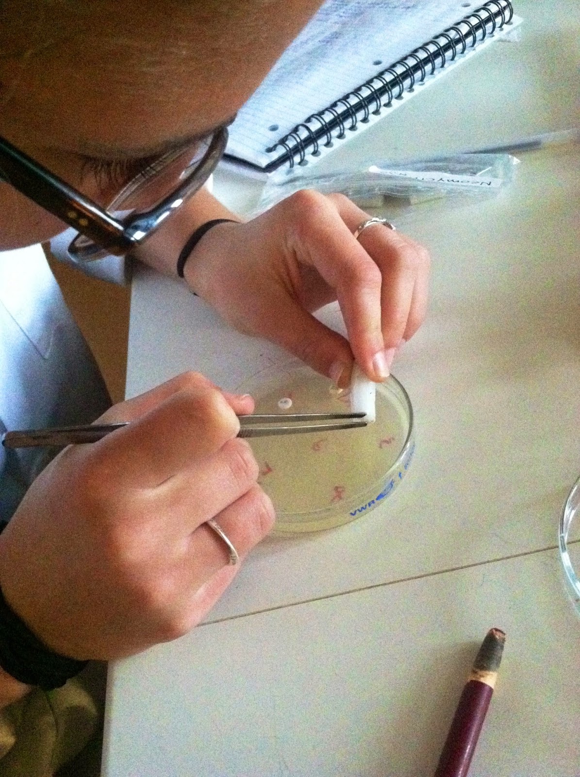
For the first three drugs we used small forceps, a small beaker filled with ethanol and a bunsen burner to sterilize the forceps.  After marking a chart of where each antibiotic was, we placed three paper discs of the drug onto the plate with the sterilized forceps (that had been dipped in the alcohol and placed over the bunsen burner flame) .


For the last three we used the same paper discs but added the antibiotic with a pipet of 2.0 micrometers.


Then we placed the inoculated plate with the antibiotics in 30 degrees Celsius incubator and eagerly wait for the results :)